Home » Without Label » Leg Bones Diagram / 239 Tibia Fibula Leg Bones Photos Free Royalty Free Stock Photos From Dreamstime : The lower leg extends from the knee to the ankle.
Leg Bones Diagram / 239 Tibia Fibula Leg Bones Photos Free Royalty Free Stock Photos From Dreamstime : The lower leg extends from the knee to the ankle.
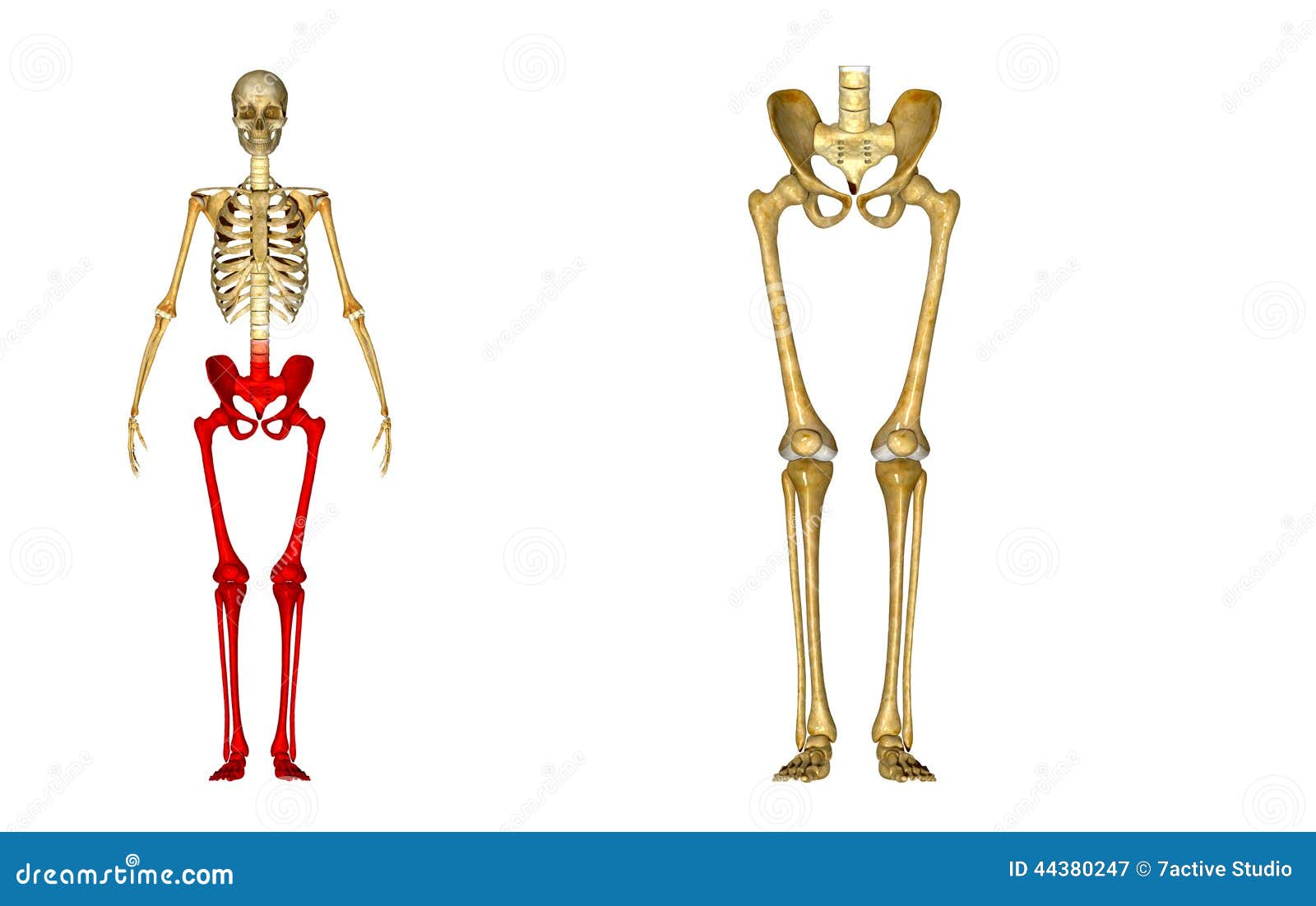
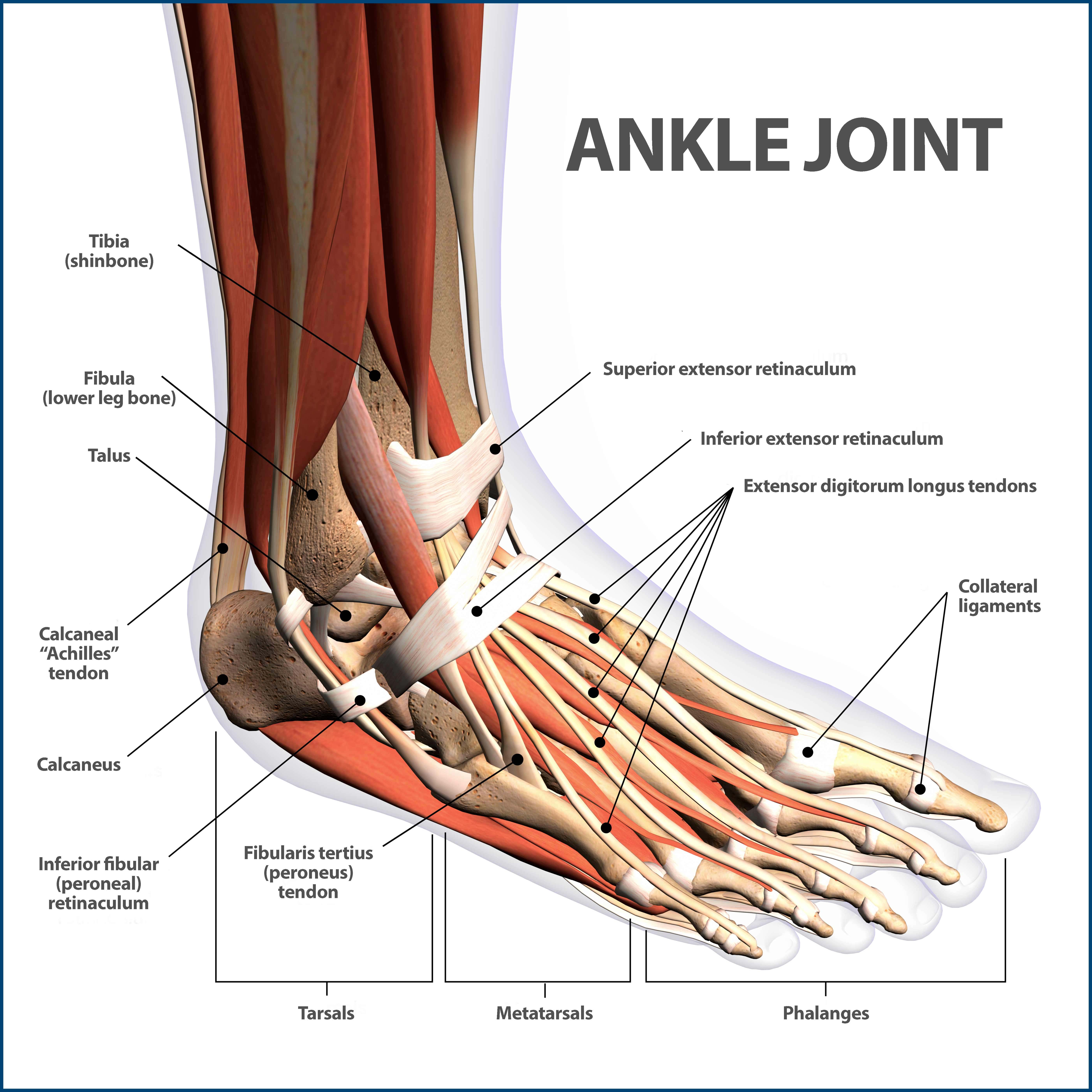
Leg Bones Diagram / 239 Tibia Fibula Leg Bones Photos Free Royalty Free Stock Photos From Dreamstime : The lower leg extends from the knee to the ankle.. The femur, or thigh bone, is the single bone of the thigh region (figure 6.51). The bones of the leg are the femur, tibia, fibula and patella. The human leg, in the general word sense, is the entire lower limb of the human body, including the foot, thigh and even the hip or gluteal region. These muscles work together to produce movements such as standing, walking, running, and jumping. The foot bones shown in this diagram are the talus, navicular, cuneiform, cuboid, metatarsals and calcaneus.
Diagram of a radious bone 12 photos of the diagram of a radious bone diagram of radius bone, bone, diagram of radius bone The medial, larger bone of the lower leg. The tibia and fibula are two long bones that run parallel to each other, forming the scaffold of the leg and providing attachment points for many muscles. Gastrocnemius muscle anatomy 17 photos of the gastrocnemius muscle anatomy deltoid muscle anatomy, gastrocnemius muscles, gracilis muscle anatomy, plantaris muscle anatomy, quadriceps muscle anatomy, sartorius muscle anatomy, soleus muscle anatomy, trapezius muscle anatomy, foot, deltoid muscle anatomy, gastrocnemius. The lower leg is comprised of two bones, the tibia and the smaller fibula.
Leg Anatomy Bones Anatomy Drawing Diagram from cdn.shopify.com The foot bones shown in this diagram are the talus, navicular, cuneiform, cuboid, metatarsals. The bone at the top of the leg. The knee joint is the largest joint in the body and is primarily a hinge joint although some sliding and rotation occur. The largest and most medial leg bone, forming both the knee and ankle joints. The proximal portion of the tibia is tibial plateau which acts as a cusp for the knee, the distal portion tapers into the medial malleoli and the concave surface which articulates with the talus at the ankle joint. The rounded, proximal end is the head of the femur, which articulates with the acetabulum of the hip bone to form the hip joint. Related posts of diagram of leg bones diagram of a radious bone. The lower leg is comprised of two bones, the tibia and the smaller fibula.
Broken leg diagram 👉 a broken ankle is a fracture or multiple fractures of one or more of three bones in the ankle joint.
The largest and most medial leg bone, forming both the knee and ankle joints. Includes leg (femur, tibia, patella, and fibula) and foot (tarsals and digits) bones. At the same time, the bones and joints of the leg and foot must be strong enough to support the body's weight while remaining. Your legs are two of your most important body parts. The knee joint is the largest joint in the body and is primarily a hinge joint, although some sliding and rotation occur. The human leg, in the general word sense, is the entire lower limb of the human body, including the foot, thigh and even the hip or gluteal region. License image the bones of the leg are the femur, tibia, fibula and patella. These muscles work together to produce movements such as standing, walking, running, and jumping. Human foot bones anatomy sketch of orthopedics medicine. These landmarks are the anterior superior iliac spine. With different grades of sprains depending on severity. The foot bones shown in this diagram are the talus, navicular, cuneiform, cuboid, metatarsals and calcaneus. The foot bones shown in this diagram are the talus, navicular, cuneiform, cuboid, metatarsals and calcaneus.
Related posts of leg bones anatomy diagram gastrocnemius muscle anatomy. The foot bones shown in this diagram are the talus, navicular, cuneiform, cuboid, metatarsals and calcaneus. These landmarks are the anterior superior iliac spine. Leg bone diagram / 3d skeletal system 5 cool facts about the femur. Bone surfaces at synovial joints are protected by a coating of articular cartilage.
239 Tibia Fibula Leg Bones Photos Free Royalty Free Stock Photos From Dreamstime from thumbs.dreamstime.com Long bones are found in the arms (humerus, ulna, radius) and legs (femur, tibia, fibula), as well as in. An atlas of cat anatomy. Diagram of a radious bone 12 photos of the diagram of a radious bone diagram of radius bone, bone, diagram of radius bone Related posts of leg bones anatomy diagram gastrocnemius muscle anatomy. He leg's main function in the human is for locomotion and support of the rest of the body. The lower leg extends from the knee to the ankle. Bone surfaces at synovial joints are protected by a coating of articular cartilage. 2006 kia optima belt diagram.
The smaller lateral bone of the lower leg.
The foot bones shown in this diagram are the talus, navicular, cuneiform, cuboid, metatarsals and calcaneus. The foot bones shown in this diagram are the talus, navicular, cuneiform, cuboid, metatarsals and calcaneus. The human leg, in the general word sense, is the entire lower limb of the human body, including the foot, thigh and even the hip or gluteal region. The lower leg is comprised of two bones, the tibia and the smaller fibula. The foot bones shown in this diagram are the talus, navicular, cuneiform, cuboid, metatarsals and calcaneus. Leg bone diagram / 3d skeletal system 5 cool facts about the femur. The bone at the top of the leg. Long bones are found in the arms (humerus, ulna, radius) and legs (femur, tibia, fibula), as well as in. The lower extremity, commonly referred to as the leg, contains four bones (the femur, the patella, the tibia, and the fibula) and bends at the hip, the knee, and the ankle. The knee joint is the largest joint in the body and is primarily a hinge joint, although some sliding and rotation occur. The bones of the leg are the femur, tibia, fibula and patella. The femur, or thigh bone, is the single bone of the thigh region (figure 6.51). The major bones of the leg are the femur (thigh bone), tibia (shin bone), and adjacent fibula, and these are all long bones.the patella (kneecap) is the sesamoid bone in front of the knee.most of the leg skeleton has bony prominences and margins that can be palpated and some serve as anatomical landmarks that define the extent of the leg.
These landmarks are the anterior superior iliac spine. The largest and most medial leg bone, forming both the knee and ankle joints. It joins with the scapula above at the shoulder joint (or. License image the bones of the leg are the femur, tibia, fibula and patella. Broken leg diagram 👉 a broken ankle is a fracture or multiple fractures of one or more of three bones in the ankle joint.
Ankle Fractures Broken Ankle Florida Orthopaedic Institute from www.floridaortho.com Its lower end helps create the knee joint. The proximal portion of the tibia is tibial plateau which acts as a cusp for the knee, the distal portion tapers into the medial malleoli and the concave surface which articulates with the talus at the ankle joint. Ankle & lower leg anatomy. The pubis, ischium, and ilium together constitute the pelvis while the thigh bone is the femur. The knee joint is the largest joint in the body and is primarily a hinge joint, although some sliding and rotation occur. The foot bones shown in this diagram are the talus, navicular, cuneiform, cuboid, metatarsals and calcaneus. The bones of the hip include the femur, the ilium, the ischium, and the pubis. The tibia and the fibula, at the top of the ankle joint.
An atlas of cat anatomy.
These muscles work together to produce movements such as standing, walking, running, and jumping. The bone at the top of the leg. Its lower end helps create the knee joint. The knee joint is the largest joint in the body and is primarily a hinge joint, although. Bone surfaces at synovial joints are protected by a coating of articular cartilage. Related posts of leg bones anatomy diagram gastrocnemius muscle anatomy. The foot bones shown in this diagram are the talus, navicular, cuneiform, cuboid, metatarsals and calcaneus. Related posts of diagram of leg bones diagram of a radious bone. The bones of the leg are the femur, tibia, fibula and patella. He leg's main function in the human is for locomotion and support of the rest of the body. Also called the shin bone, the tibia is the longer of the two bones in the. The tibia and the fibula, at the top of the ankle joint. The proximal portion of the tibia is tibial plateau which acts as a cusp for the knee, the distal portion tapers into the medial malleoli and the concave surface which articulates with the talus at the ankle joint.